Synopsis
When a shoulder, hip, or ankle injury strikes, patients are often forced into a waiting loop for an expensive, static MRI to pinpoint the damage. This guide explores the clinical shift toward dynamic diagnostic ultrasound—a highly precise imaging modality that allows specialists to view your muscles, tendons, and ligaments in motion. We detail how the integrated orthopaedic model at Orthocure Clinics and Strength Studios utilizes real-time ultrasound imaging to catch subtle soft-tissue tears and joint impingements that traditional, stationary scans often miss. By obtaining an immediate, accurate look at your tissue biology, we can seamlessly design a targeted path for structural correction. We examine how our specialized physiotherapy team translates these precise visual findings into customized, low-impact loading strategies. Understanding exactly what is happening beneath your skin is the first step toward fast, definitive joint pain relief. Consistent application of these diagnostic insights within our Strength Studio ensures you train safely, protecting your healing fibers while building an internal muscular shield against future injury.
Table of Contents
- Moving Beyond the Static Scan- What Is Dynamic Diagnostic Ultrasound?
- The Real-Time Advantage- Watching Tendons and Ligaments in Motion
- Catching the Silent Tears- Rotator Cuff, Plantar Fascia, and Tendon Patches
- Ultrasound vs. MRI- A Comparative Look at Imaging Physics
- Structural Correction- Aligning the Framework Based on Visual Data
- Specialized Physiotherapy- Using Live Scans to Guide Hands-On Care
- The Strength Studio- Calibrated Loading for Validated Tissue Healing
- Partnering with Orthocure for Diagnostic Precision and Rapid Recovery
Moving Beyond the Static Scan- What Is Diagnostic Ultrasound?
For decades, standard orthopaedic diagnostics relied almost entirely on X-rays and MRIs. While an X-ray captures bone and an MRI provides a highly detailed image of deep internal anatomy, they both share a major limitation: they are completely static. At Orthocure Clinics and Strength Studios, we utilize musculoskeletal diagnostic ultrasound to change this narrative. By using high-frequency sound waves, this technology allows our clinicians to peer directly beneath the skin to evaluate the architecture of your soft tissues while you are sitting, standing, or actively moving.
The Real-Time Advantage- Watching Tendons and Ligaments in Motion
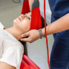
The true magic of diagnostic ultrasound lies in its dynamic capability. Many soft-tissue problems only occur during a specific movement, such as when a rotator cuff tendon pinches against a bone at a certain angle of shoulder elevation. During an MRI, a patient lies completely still, which can cause these functional impingements to remain completely hidden. An ultrasound allows a specialist to actively move your joint, press on the exact point of discomfort, and watch the tendons glide, contract, or catch in real time.
Catching the Silent Tears- Rotator Cuff, Plantar Fascia, and Tendon Patches
Dynamic ultrasound is exceptionally sensitive when mapping superficial soft tissues. It allows our team to easily identify micro-tears in the tendons of the shoulder, tennis elbow strains in the forearm, chronic thickening of the plantar fascia, and acute ligament sprains in the ankle. Because we can measure the exact width and depth of a tear in millimeters, we remove the guesswork from your prognosis. This clarity is essential for mapping out a reliable path toward long-term joint pain relief .
Ultrasound vs. MRI- A Comparative Look at Imaging Physics
To understand why this modality has become an indispensable first line of defense, it helps to see how it compares directly to an MRI scan:
Imaging Feature | Diagnostic Ultrasound | Magnetic Resonance Imaging (MRI) |
|---|---|---|
Tissue State | Dynamic; evaluated during active movement. | Static; requires total immobility inside a tube. |
Metal Implants | Completely unaffected; can scan directly over hardware. | Distorted; metal causes severe visual artifacts. |
Claustrophobia | Zero; performed in an open, comfortable clinic room. | High; can be stressful for claustrophobic patients. |
Primary Use Case | Superficial tendons, ligaments, nerves, and muscles. | Deep intra-articular structures, bone marrow, and discs. |
Structural Correction- Aligning the Framework Based on Visual Data
An accurate diagnosis dictates the success of your recovery. If an ultrasound reveals that a shoulder tendon is tracking poorly due to a slumped upper back, attempting to treat the arm locally will fail. Structural correction at Orthocure uses these live imaging insights to correct your overall skeletal framework. By realigning a rotated pelvis or normalizing a forward pelvic tilt , we change the angle at which your muscles pull, immediately taking the mechanical strain off the partially torn or inflamed tissue.
Specialized Physiotherapy- Using Live Scans to Guide Hands-On Care
Our specialized physiotherapy protocols are directly informed by your live tissue scans. If the ultrasound reveals a localized pocket of fluid (bursitis) or disorganized scar tissue from an old injury, our physiotherapists customize their manual therapy techniques with surgical precision. We know exactly where to apply targeted myofascial release, dry needling, or gentle joint distraction to stimulate local blood circulation and promote healthy, parallel collagen remodeling.
The Strength Studio- Calibrated Loading for Validated Tissue Healing
Once the exact boundary of a soft-tissue tear is known, rehabilitation can progress safely into our Strength Studio . Inside our medical-grade Medical Gym environment, we use highly isolated resistance training to load the healing tissue without risking further damage. Because we know the structural limit of the tendon, we can prescribe precise weights and angles that stimulate cellular repair without exceeding the tissue’s current tolerance. This data-driven precision ensures your recovery is fast, efficient, and completely secure.
Partnering with Orthocure for Diagnostic Precision and Rapid Recovery
Do not waste weeks guessing the source of your muscle or joint pain. Our comprehensive services at Orthocure Clinics and Strength Studios integrate real-time diagnostic imaging with cutting-edge physical medicine. By combining dynamic ultrasound imaging, targeted structural correction, specialized manual therapy, and medical-grade conditioning, we provide a smooth, non-surgical pathway to full recovery. Partner with Orthocure today and see your injury clearly to fix it permanently.
FAQs
Does a diagnostic ultrasound expose my body to harmful radiation?
No, absolutely not. Unlike X-rays or CT scans, diagnostic ultrasound uses zero ionizing radiation. It relies entirely on high-frequency sound waves that bounce off internal tissues to create a live visual image. It is exceptionally safe, completely painless, and can be used repeatedly to monitor your tissue healing progress.
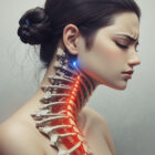
Can an ultrasound scan see inside my spinal column to find a herniated disc?
No. High-frequency sound waves cannot easily penetrate dense, solid bone structures like the vertebrae of your spine. While ultrasound is the absolute gold standard for scanning superficial joints, muscles, and tendons (like your shoulder, elbow, or ankle), evaluating deep spinal cords, bones, or a suspected lumbar disc herniation still requires an MRI.
Why is a dynamic ultrasound scan often preferred over a standard MRI for a shoulder injury?
A shoulder is a highly mobile joint. An MRI captures a beautiful, high-resolution snapshot, but it can miss a “functional impingement”—a situation where a bone only scrapes a tendon when you actively raise your arm. By moving your shoulder during a diagnostic ultrasound , we can witness the exact millisecond the tissue hitches, leading to a much more accurate diagnosis.
Is it safe to perform heavy exercises in the Medical Gym if I have a partial tendon tear?
It is safe only if the exercise is precisely calibrated. In our Medical Gym , we use the visual blueprints from your ultrasound to design a “safe zone” for movement. We avoid ballistic stretches that could expand the tear, focusing instead on slow, isolated isometric and eccentric loading in our Strength Studio to stimulate fresh collagen alignment.
How long does a diagnostic ultrasound screening take at the clinic?
A focused musculoskeletal ultrasound evaluation typically takes between 15 to 30 minutes. During this time, our orthopaedic specialist will scan the injured area, guide you through various movements to observe the tissue glide, explain the visual findings on the monitor, and immediately coordinate your treatment plan with our specialized physiotherapy team.